Metamorphosis and Development
Kelsey Martin
Apoptosis in Ontogeny
The growth and development of an organism from conception to maturity is called ontogenesis (free dictionary.com). Development of an organism involves many processes and signals to progress the embryo to the mature, adult form. The developmental process is normally associated with proliferative events, however apoptosis plays a significant role in development as well.The involvement of apoptosis in ontogeny was first discovered in the 1950s. Glucksmann et al. had observed that cell death occurred during the embryonic development in many mammals. They then concluded that cell death was an intentional part of development. (Perez-Garijo et al., 2013). "Cell death via apoptosis is as much a part of embryonic development as cell proliferation is for differentiation" (Haanen et al., 1996). One helpful analogy for understanding apoptosis in development is to compare it to the sculpting a marble statue. A sculptor must carve off pieces of the block of marble to achieve the final statue. Apoptosis is used in the same way during development to achieve the final morphology (Haanen et al., 1996).
One of the first ways developmental apoptosis was identified was through studying the common fruit fly, Drosophila melanogaster. Figure 1 displays the pathway of apoptosis in the fly. When development occurs, developmental proteins such as Hox activate apoptotic signaling proteins, such as Reaper, Grim, and Hid. These proteins inhibit DIAP1, which is an inhibitor of apoptosis. Normally, DIAP1 inhibits the formation of Dronc, which is a protein with caspase-9 qualities. When DIAP1 is inhibited though, Dronc is active in the cell. Dronc can then activate DrICE and Dcp-1, which exhibit caspase-3 like qualities. DrICE and Dcp-1 then induce apoptosis.
When the apoptotic proteins Grim, Reaper, and Hid are inhibited, apoptosis cannot occur. Inhibition of these apoptotic proteins has been linked to fly lethality and severe developmental defects can occur. (Fuchs et al., 2011) The results from Fuchs et al. indicated the importance of cell death in the development of the fruit fly.
Figure 1 shows the apoptotic pathways of not only the common fruit fly, but of C. elegans and mammals. C. elegans, or the nematode, is a simple multi-cellular organism that contains a simple apoptotic pathway. Mammals exhibit a more complex apoptotic pathway. The figure shows the increasing complexity of apoptotic pathways as organisms increase in their complexity as well.
 |
| Figure 1. Apoptotic pathways in C. elegans, Drosophila, and mammals. From simple to complex, the organisms exhibit increased complex apoptotic pathways. |
Functions of Apoptosis in Development
Apoptosis is important (and necessary) in the development of organisms. There are four main ways that apoptosis affects development. The first way apoptosis affects development is through the sculpting of structures, such as digits. Apoptosis is used to delete structures unnecessary for the mature organism. Apoptosis is also used to control cell number in the organism, and finally apoptosis is used to kill harmful cells. All four of these examples are illustrated in figure 2.
 |
| Figure 2. The four main ways apoptosis regulates development. |
1. Sculpting Structures
When an organism develops, cell death is required to help sculpt certain organs or features. One example is the degeneration of interdigital tissue, such as in mammalian paws and hands. (Suzanne et al., 2013).
For the separation of digits, the genes Bmp2 and Bmp4 are expressed in the interdigital regions. Figure 3 shows the pathway for how BMP induces apoptosis.
 |
| Figure 3. Zhang et al. 2003 Pathway for BMP induced apoptosis in interdigital regions in organisms. |
In the above pathway, when BMP (bone morphogenetic protein) binds to receptors I and II, the receptors heterodimerize and form a receptor-ligand complex. Receptor I is phosphorylated and subsequently activated by receptor II. The activated receptor I then phosphorylates and activates Receptor-activated Smads (R-Smads), such as Smad1. Activated R-Smads can then bind with Co-Smads such as Smad4 and can enter the nucleus and down regulate the transcription of bcl-2. (Zhang et al., 2003).
Bcl-2 is an anti-apoptotic gene that when present in the cell blocks cytochrome c release into the cytoplasm and increases cytosolic potassium concentration as well. When bcl-2 is down regulated though, there is an increase in cytosolic cytochrome c concentration. The increase of cytochrome c activates caspase-3, leading to apoptosis. The down regulation of bcl-2 also leads to a decrease of potassium, which in turn leads to a decrease of apoptotic volume. The decrease of apoptotic volume leads to an increase in apoptosis (Zhang et al., 2003).
Lindsten et al. in 2000 wanted to see how the removal of several pro-apoptotic genes in mice affected the phenotype of their paws. Bax and Bak are both Bcl-2 family pro-apoptotic genes. As mentioned in the explanation of figure 3, Bcl-2 is also an anti-apoptotic gene. Different families of Bcl-2 can either be anti or pro-apoptotic. Bax and Bak are both pro-apoptotic Bcl-2 genes that when activated, promote cytochrome c release. As seen in the pathway above, increased cytochrome c concentration leads to an increase in apoptosis.
When bax and bak were completely knocked out, there was a severe digit webbing phenotype observed that was not observed in the heterozygous knockout, as seen in figure 4. When complete homozygous mutant mice were bred, less than 10% of the mutants survived. The reduced birth rate in the homozygous mutants led Lindsten et al. to the assumption that homozygous knockout of the Bcl-2 family genes caused greater instances of embryonic lethality.
 |
| Figure 4. Anatomical phenotypes of bax bak mice. (a)(c) bax+/-bak-/- paws (b)(d) bax-/- bak -/- paws. When there is a complete homozygous knockout of both of these genes, severe webbing is seen. |
Lohmann et al. wanted to see how Dfd affected head development. The Dfd gene was knocked out in the fly, and the levels of apoptotic proteins were compared. Compared to wild-type, there was a significant decrease in the number of apoptotic cells in Dfd knockout flies. These results suggest that the hox protein Dfd regulates apoptosis in the fly. Expression of common pro-apoptotic genes was then compared in the segments, and reaper specifically was significantly expressed at segmental boundaries (Lohmann et al., 2002).
Lohmann et al. then wanted to compare wild-type flies with Dfd mutant flies in head morphology. As seen in figure 5, there are three main head segments- maxillary (mx), mandibular (md), and labial (lb). Figure E shows an enlarged picture from figure B and displays the wild-type phenotype. The three three head segments have distinct boundaries and are separated from each other. Homozygous Dfd mutant flies show no distinct separation, especially in the mandibular and maxillary head segments, as seen in figure G. (Lohmann et al., 2002). These results suggest that apoptosis is important in head development in the fly.
 |
| Figure 5. Head morphology in Dfd mutant fruit flies. Figures B and E shows wild-type head morphology. Figures C and F shows head morphology for heterozygous Dfd mutants. Figures D and G show head morphology for homozygous Dfd mutants. |
2. Deletion of Structures
Another way apoptosis is involved in development is through the deletion of certain structures. In organisms, certain structures are sex specific. Once sex determination has occurred during the early stages of development, certain structures are no longer necessary and end up being deleted. In mammals, two ducts exist- the Müllerian duct and the Wolffian duct. In female mammals, the Müllerian duct develops into the ovaries and the uterus. In males, the Wolffian duct forms the vas deferens, epididymis, and seminar vesicle. Once sex determination has occurred, the unnecessary duct is slowly eliminated via apoptotic cells. Female mammals delete the Wolffian duct, and males delete the Müllerian duct.
(Fuchs et al., 2011).
Click here to see a video of the formation of the female reproductive system via apoptosis of the Wolffian duct.
3. Regulating Cell Number
Another role of apoptosis in development is through the regulation of cell number. One way apoptosis regulates cell number in development is through the regulation of oocytes. Early on in development, more than 99.9% of ovarian follicles undergo a degenerative process that leads to follicle atresia (Haanen et al., 1995). Follicle atresia is the natural breakdown of oocytes throughout a female's lifespan. Human females are born with millions of oocyte follicles in the ovaries when born. However, due to follicle atresia, only around 400 oocytes are ovulated (Haanen et al., 1995).
Follicle atresia is regulated by granular apoptosis, and consists of several "death" proteins that are continuously activated leading to follicle atresia:
TNF-alpha, Fas ligand, TRAIL (TNF-related apoptosis-inducing ligand), and APO-3 ligand (Inoue et al., 2003).
According to Inoue et al., 2003, TRAIL activates caspase-3, which induces apoptosis in the oocyte follicles. Follicle stimulating hormone (FSH) inhibits the pro-apoptotic proteins.
4. Elimination of abnormal cells
 |
| Figure 6 . Positive and negative selection for T cells. |
Apoptosis in Metamorphosis
 |
| http://www.revuemag.com/2011/06/metamorphosis/ |
Not only is apoptosis important in development, apoptosis plays a crucial role in metamorphosis. Metamorphosis is "a biological process in which an animal physically develops after birth, involving a conspicuous and abrupt change in the animal's form or structure through cell growth and differentiation" (biology-online.org). Metamorphosis is commonly associated with the Monarch butterfly and amphibians such as frogs. For amphibian metamorphosis, the tadpole changes from its aquatic form into a terrestrial form, the frog. Apoptosis is an important mechanism in this transition (Atsuko et al., 2010).
From Youtube.
TUNEL assay
 |
| https://www.rndsystems.com/products/tdt-in-situ-apoptosis-detection-kit--dab_4810-30-k
TUNEL assay, or terminal deoxynucleotidyl transferase dUTP Nick End Labeling assay, is used to identify if apoptosis is occurring in the cell. According to Jena Bioscience, labeled dUTPS attach to the 3'-OH end of DNA strand breaks. The fluorescence is visualized by the label attached to the dUTPs.
|
Amphibian Metamorphosis
During metamorphosis of the tadpole, thyroid hormone levels were measured at different stages. As seen in figure 7, there was a spike in the plasma thyroid hormone level during tail cell death. They also discovered that most apoptotic events occur during the climax of the thyroid hormone spike. The discovery led researchers to the conclusion that thyroid hormone regulates apoptosis (Atsuko et al., 2012).
Atsuko et al. also noted that other developmental events, such as the notochord and spinal cord apoptosis, do not occur exactly when tail apoptosis occurs. However, they occur during the metamorphic climax and are influenced by the presence of thyroid hormone.
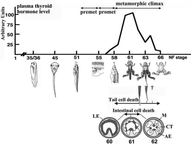 |
Figure 7. The plasma thyroid hormone levels during different stages of metamorphosis.
|
Thyroid Hormone Apoptotic Pathway
The thyroid hormone apoptotic pathway used in tadpole metamorphosis is displayed in figure 8. Thyroid Hormone, or T3, binds to a thyroid hormone receptor. The binding activity activates and up-regulates Bax, a pro-apoptotic protein. Bax then locates to the surface of the mitochondria and directly activates cytochrome c release into the cytoplasm. Cytochrome c then binds and forms a complex with APAF-1, which stands for apoptotic protease activating factor 1. The complex then activates caspase-9, which in turn activates caspase-3. Activated caspase-3 activates apoptosis and causes cell death in the tadpole to induce metamorphosis. (Rowe et al., 2005)
 |
| Figure 8. The apoptotic pathway of T3 in amphibian metamorphosis. |
Conclusion
Overall, apoptosis is involved in many developmental and metamorphic processes. Programmed cell death is observed in many processes, such as in the sculpting of digits in mice and in the metamorphosis of tadpoles. With these examples, one can see the importance of cell death in sustaining life.
Hopefully this page has helped better your understanding of apoptosis and its role in development and metamorphosis.

No comments:
Post a Comment